Bones In Leg Diagram - Bones Of The Human Leg And Foot Scienceaid - Leg diagram bones posted on july 16, 2018 by admin human leg anatomy bones stock images royalty free vectors shutterstock physiology anatomy of bones in leg skeletal series part 10 the human these mine lower leg bones anatomy bone human body leg bone anatomy hd images download.
byAdmin•
0
Bones In Leg Diagram - Bones Of The Human Leg And Foot Scienceaid - Leg diagram bones posted on july 16, 2018 by admin human leg anatomy bones stock images royalty free vectors shutterstock physiology anatomy of bones in leg skeletal series part 10 the human these mine lower leg bones anatomy bone human body leg bone anatomy hd images download.. When your muscles contract, they pull the bone they're. Editor · aug 13, 2017 ·. The humerus and the femur are corresponding bones of the arms and legs, respectively. 11.02 x 15.75 in (unframed). Human anatomy diagrams show internal organs, cells, systems, conditions, symptoms and sickness information and/or tips for healthy living.
This condition happens when the spaces within the bones in your spine get narrow. The bones of the leg are the femur, tibia, fibula and patella. Pencil drawing on paper one of a kind artwork size: This lengthy bone connects with the knee at one finish and the ankle on the different. Nervsystemet anatomy, diagram & function | health.
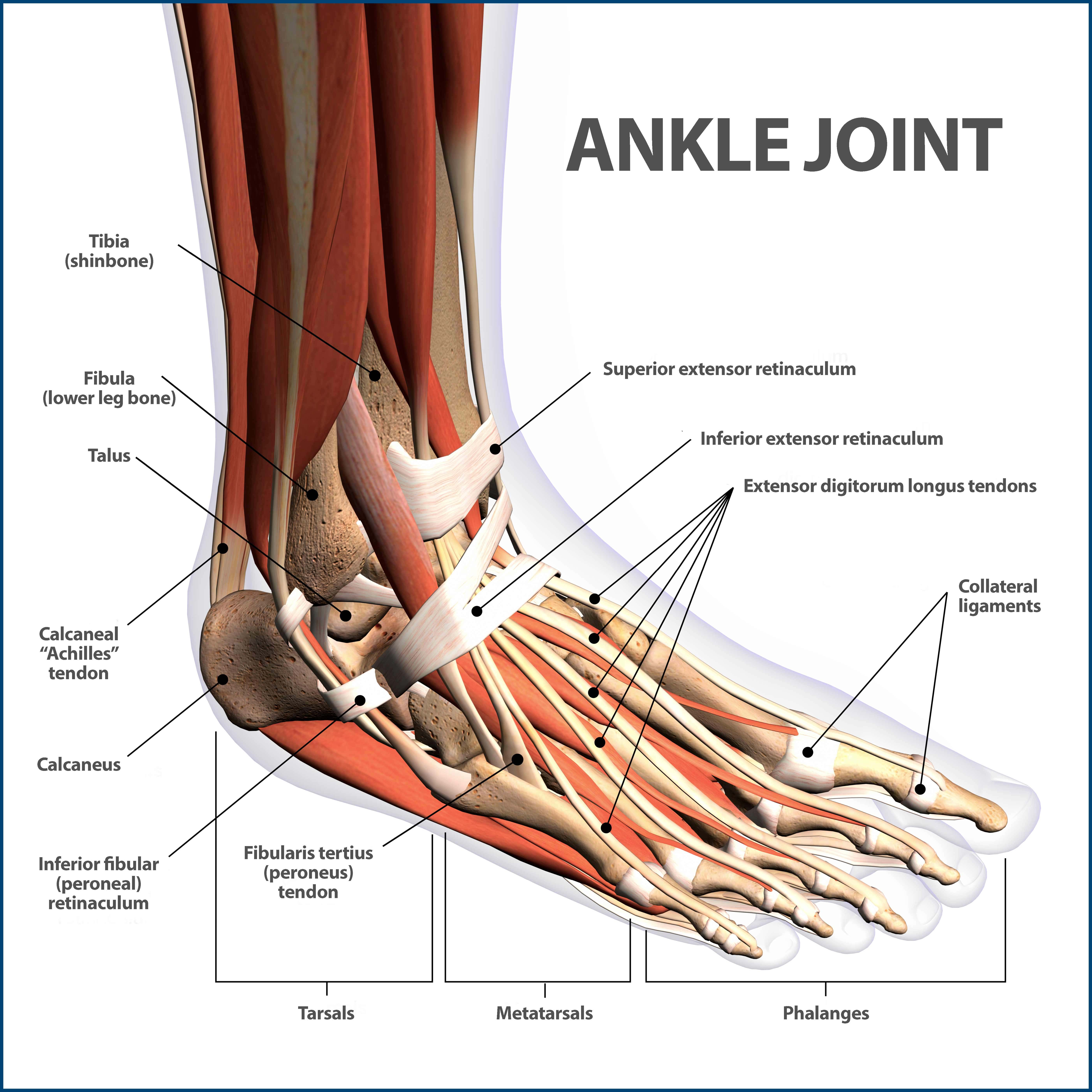
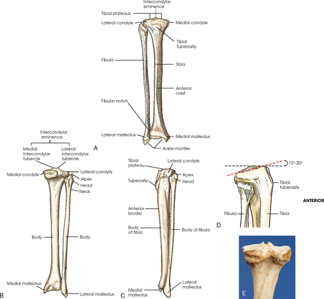
Ankle Fractures Broken Ankle Florida Orthopaedic Institute from www.floridaortho.com Mcqs on leg bones for neet. Top suggestions for human leg bones diagram. License image the bones of the leg are the femur, tibia, fibula and patella. When your muscles contract, they pull the bone they're. The humerus and the femur are corresponding bones of the arms and legs, respectively. The foot bones shown in this diagram are the talus, navicular, cuneiform, cuboid, metatarsals and calcaneus. At the distal end of the femur, two rounded condyles meet the tibia and fibula bones of the lower leg to form the knee joint. This diagram of a human skeleton is labeled with 12 major bones, from skull to fibula.
That puts pressure on the nerves in the area and can cause pain, tingling.
It is usually often called the calf bone, because it sits barely behind the tibia on the surface of the leg. Top suggestions for human leg bones diagram. A small sesamoid bone in the knee joint called patella. But if something goes a bit wrong, they can hurt and make it hard to move around. The foot bones shown in this diagram are the talus, navicular, cuneiform, cuboid, metatarsals and calcaneus. That puts pressure on the nerves in the area and can cause pain, tingling. An electrical wiring diagram can be as simple as a diagram demonstrating how to set up a fresh swap with your hallway. This condition happens when the spaces within the bones in your spine get narrow. 11.02 x 15.75 in (unframed). The foot bones shown in this diagram are the talus, navicular, cuneiform, cuboid, metatarsals and calcaneus. Editor · aug 13, 2017 ·. Human anatomy diagrams show internal organs, cells, systems, conditions, symptoms and sickness information and/or tips for healthy living. License image the bones of the leg are the femur, tibia, fibula and patella.
Your legs are an amazing collection of bones and muscles. The bones of the leg are the femur, tibia, fibula and patella. The bones of the leg are the femur, tibia, fibula and patella. What does this suggest about mammals? Continue scrolling to read more below.
Bone Fracture Human Leg Anatomy And Skeleton Stock Vector Illustration Of Medicine Barefoot 114048504 from thumbs.dreamstime.com Most of the animals have the same bones, although some are shaped differently and placed in different positions. Want to learn more about it? The lower limb has 30 bones some of which are tibia, femur, tarsal bones, fibula, metatarsal bones, etc. The foot bones shown in this diagram are the talus, navicular, cuneiform, cuboid, metatarsals and calcaneus. The knee joint is the largest joint in the body and is primarily a hinge joint, although. When your muscles contract, they pull the bone they're. Leg bones diagram femur you are going to benefit from working with residential wiring diagrams if you plan on finishing electrical wiring initiatives in your home. It is unique in that it's particularly designed to be wound as limited as possible to make a potent magnetic area and is likewise enveloped in a thin layer of insulation.
The humerus and the femur are corresponding bones of the arms and legs, respectively.
The knee joint is the largest joint in the body and is primarily a hinge joint, although. Download this free vector about diagram showing the hip bone treatment, and discover more than 15 million professional graphic resources on freepik. Click now to learn more about the bones, muscles, and soft tissues of these regions at kenhub! Traditional methods to evaluate stress may be stressful to the bird. A small sesamoid bone in the knee joint called patella. Color the leg on the left side. When you stand or walk, all the weight of your upper body rests on them. Master leg and knee anatomy using our topic page. Human anatomy diagrams and charts show internal organs, body systems, cells, conditions, sickness and symptoms information and/or tips to ensure one lives in good health. The human leg, in the general word sense, is the entire lower limb of the human body, including the foot, thigh and even the hip or gluteal region. D) that the shape of the bones has less to do with the environment. Most of the animals have the same bones, although some are shaped differently and placed in different positions. Tibia and fibula, the leg bones.
The lower limb has 30 bones some of which are tibia, femur, tarsal bones, fibula, metatarsal bones, etc. Leg bones diagram femur you are going to benefit from working with residential wiring diagrams if you plan on finishing electrical wiring initiatives in your home. When your muscles contract, they pull the bone they're. Mcqs on leg bones for neet. The humerus and the femur are corresponding bones of the arms and legs, respectively.
Lower Limb Radiology Key from radiologykey.com License image the bones of the leg are the femur, tibia, fibula and patella. This diagram of a human skeleton is labeled with 12 major bones, from skull to fibula. Human anatomy diagrams show internal organs, cells, systems, conditions, symptoms and sickness information and/or tips for healthy living. Learn vocabulary, terms and more with flashcards, games and other study tools. Muscles and bones of the upper leg and pelvis; Editor · aug 13, 2017 ·. Continue scrolling to read more below. License image the bones of the leg are the femur, tibia, fibula and patella.
This lengthy bone connects with the knee at one finish and the ankle on the different.
Master leg and knee anatomy using our topic page. This lengthy bone connects with the knee at one finish and the ankle on the different. This diagram of a human skeleton is labeled with 12 major bones, from skull to fibula. The bones of your leg have roughened patches on their surfaces where muscles are attached. While their parts are similar in general, their structure has been adapted to differing functions. When your muscles contract, they pull the bone they're. The humerus and the femur are corresponding bones of the arms and legs, respectively. 11.02 x 15.75 in (unframed). The foot bones shown in this diagram are the talus, navicular, cuneiform, cuboid, metatarsals and calcaneus. The knee is a strong but flexible hinge joint. This human anatomy diagram with labels depicts and explains the details and or parts of the bones in your legs. C) that they developed their bone structure independently of one another. That puts pressure on the nerves in the area and can cause pain, tingling.